Carpal Bones Anatomy
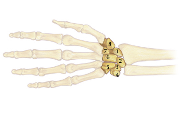
- The carpal bones are a set of eight small bones that form the wrist (or carpus), connecting the hand to the forearm. They are arranged in two rows, a proximal row and a distal row.
- Proximal row carpal bones include the scaphoid, the lunate, the triquetrum, and the pisiform.
- Distal row carpal bones include the trapezium, the trapezoid, the capitate, the hamate.
- The carpal bones are part of the radiocarpal joint, the intercarpal joints, the midcarpal joint and the CMC joints
- The dorsal ligaments are wider and thinner than the volar ligaments.
- The major dorsal carpal ligaments are the dorsal intercarpal ligament and the dorsal radiotriquetral ligament.
- The major volar carpal ligaments are the radioscaphocapitate ligament, the radioscapholunate ligament, the radiolunotriquetral ligament, the scaphotrapezial ligament, the ulnolunate ligament, and the ulnotriquetral ligament.
- The major interosseous carpal ligaments are the scapholunate ligament and the lunotriquetral ligament.
Diagrams & Photos
Key Points
- The carpal bones and the transverse carpal ligament make up the carpal bone structure.
- No tendons insert onto the carpal bones.
- The space of Poirier is a weak area in the volar wrist capsule.
- As a child grows the carpal bones ossify at different times. The last carpal bone to ossify is the pisiform at approximately 12 years of age.
