Distal Radius Anatomy
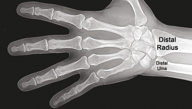
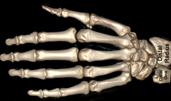
- Distal radius articulates primarily with scaphoid and lunate but also with the distal ulnar head.
- The distal radius had three facets: scaphoid facet, lunate facet, and the sigmoid notch.
- The radiocarpal joint allows flexion/extension; ulnar and radial deviation.
- Sigmoid notch is a small concavity on the medial side of the distal radius where the head of the ulna articulates.
- Rotation (supination and pronation) occurs via the distal radioulnar joint which is the joint between the ulnar head and the sigmoid notch.
- Ulnarly the distal radius and the ulna attach to the TFCC. The TFCC articulates with the triquetrum distally and the ulnar head proximally.
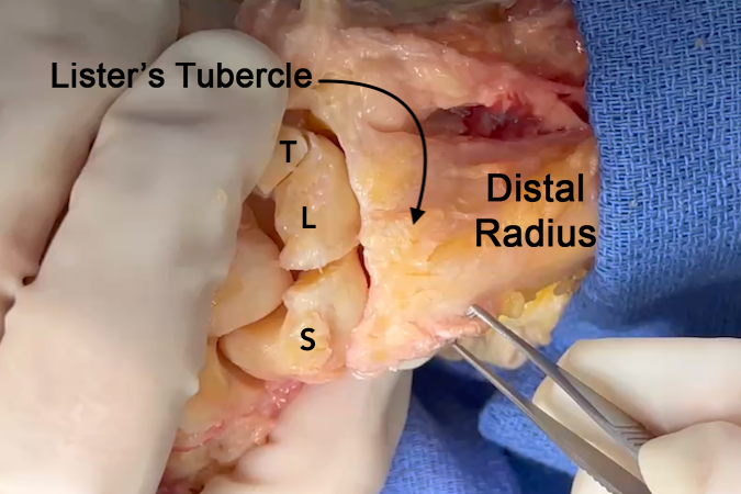
- Bony surface landmarks on the distal radius include the radial styloid with its dorsal groove for the EBP and APL and the Lister’s tubercle dorsally which provides a pivot point for the EPL.
- The radial styloid process is a bony prominence located on the lateral (radial) aspect of the radius and serves as an attachment point for ligaments that connect to the radius to the carpal bones
Diagrams & Photos
Key Points
- Palmarly the distal radius attaches to the carpus via the radioscaphocapitate ligament and the long and short radiolunate ligaments.
- Dorsally the distal radius attaches to the carpus with the radioscaphoid ligament, the radiotriquetral ligament, and dorsal capsule.
- Carpal bones of the first carpal row are interconnected by the interosseous (intrinsic) ligaments; the scapholunate and lunotriquetral ligaments.


