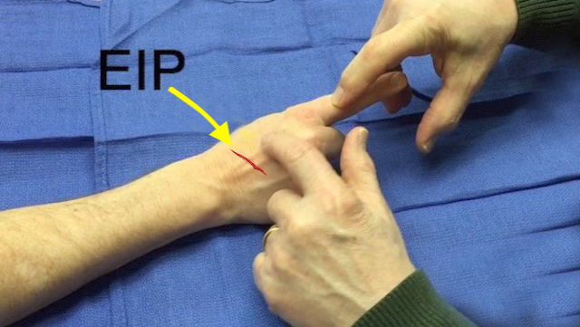
Extensor Indicis Proprius (EIP) Exam
Origin: Ulna (posterior surface of shaft) Interosseous membrane
Insertion: 2nd digit (via tendon of extensor digitorum into extensor hood)
Innervation: Cervical root(s): C7 and C8 Nerve: radial nerve (posterior interosseous branch)
EIP Examination:To test whether the EIP tendon is intact or lacerated, have patient flex the long, ring, and little fingers into the palm while actively extending the index finger. By flexing the long, ring, and little fingers the examiner isolates the function of the EIP and removes the extension forces normally provided by the EDC which can also extend the index MP joint. The purpose of the laceration exam is to determine if the tendon is intact or lacerated completely. Sometimes observing active motion or gently resisted active motion will also suggest a partial laceration.
- The brain has independent control over the function of the extensor indicis proprius (EIP) which allows the index finger to be actively extended without extending the long, ring or little fingers.
- This independent function also makes the EIP tendon an excellent motor for tendon transfers.
- The EIP tendon is often transferred to the thumb to reconstruct the function of a chronic EPL tendon laceration.
