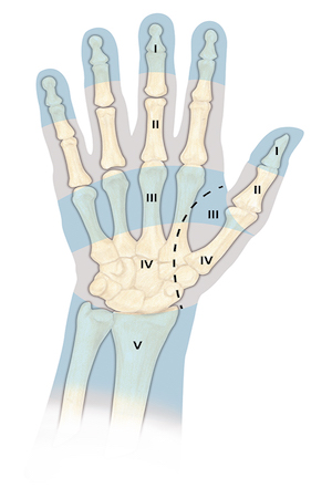
Flexor Digitorum Superficialis Exam Section 9
Flexor Digitorum Sublimis (FDS)
- Origin: Humeral-ulnar head: Humerus (medial epicondyle via the common flexor tendon), ulnar collateral ligament (UCL) of the elbow joint, ulna (coronoid process, medial side), and intermuscular septa. Radial Head: Radius (oblique line on anterior surface).
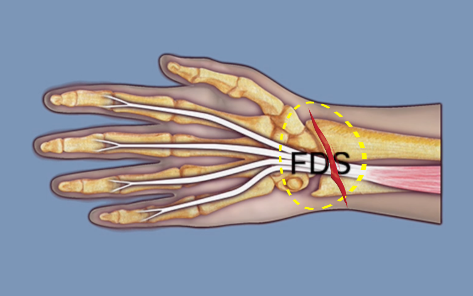
- Insertion: Four tendons arranged in two pairs: Superficial pair: Long and ring fingers, and deep Pair: Index and little fingers. All FDS tendons insert into the proximal middle phalanx.
- Innervation: Cervical root(s): C8–T1; Nerve: median nerve.
- In the proximal forearm the median nerve is on the dorsal surface of the FDS.
- The proximal flexor digitorum superficialis (FDS) has ulnar and radial heads.
- Proximally FDS muscle gets blood supply from the radial and ulnar arteries.
FDS Muscle and Tendon Testing
When assessing a finger injury for signs of flexor digitorum superficalis (FDS) laceration, the goal is to determine whether the FDS tendon is completely lacerated, partially transected, or intact.
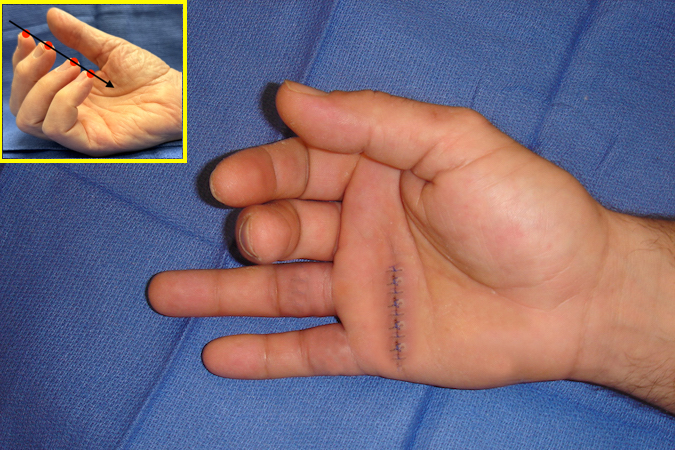
The first step in the examination is to observe the finger flexion cascade. When the normal hand is resting on the exam table with the forearm supinated and the wrist in mild dorsal flexion, the fingers are typically in a flexed position with the index minimally flexed and the little finger flexed the most. If a flexor tendon(s), FDP, FDS, or both are cut, invariably the injured finger falls out of the normal finger flexion cascade posture. If both the FDP and FDS tendons are completely cut, the finger often rests in an extended position. If a complete laceration of the FDP tendon is present, then a partially extended finger posture with DIP in extension and the PIP and MP joints flexed may be observed. Partial lacerations of either flexor tendon (FDP or FDS) may also disrupt the normal finger flexion cascade. A complete laceration of the FDS with an intact FDP could theoretically decrease the flexed posture of the involved finger but this is rare and may not disrupt the finger flexion cascade enough to be recognized.
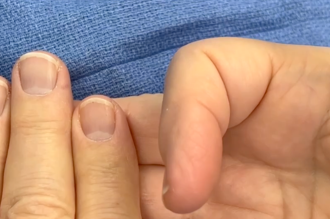
If the patient can cooperate with the request for active flexion, then the second step is to ask the patient to actively make a gentle fist. Observing active flexion of the PIP joint and the DIP joint will help identify the normal function of the FDS and FDP flexor tendons respectively.
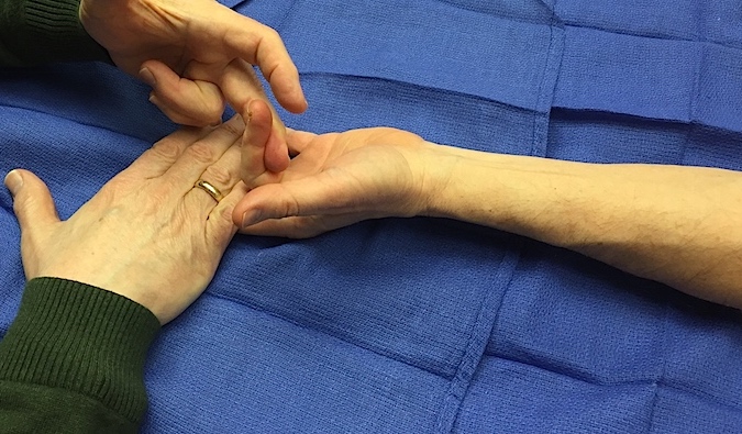
The third step is to perform muscle testing for the flexor tendons if possible. In the examination of an uninjured FDS musculotendinous unit, the 0 to 5 muscle testing grading system is applied. In this system, zero indicates a total loss of flexor digitorum profundus (FDS) contraction, while a grade of 5 represents normal FDS function capable of contracting against standard resistance. Detailed information on graded muscle testing is provided below. Typically, full muscle testing is impractical in cases of acute laceration due to pain and tenderness. The examiner may have to rely on the observation that the laceration occurred in a particular palmar section containing the FDS. Nevertheless, the examiner should assess the contraction of the potentially injured musculotendinous unit as comprehensively as possible. The examination's primary aim is to preoperatively determine whether the tendon is completely, partially cut, or intact. To evaluate the FDS, position the patient's hand and upper extremity with the forearm in supination and the wrist in a neutral position, with the hand resting on the table. Start with the fingers in a neutral resting posture. Stabilize the proximal phalanx in slight extension with one hand while resisting flexion of the PIP joint with other hand. Instruct for the patient to “Bend your finger. Hold it. Don’t let me straighten it.”
Definition of Positive Result in FDS Muscle Testing: A normal result is a positive one. During a normal muscle test, the examiner should observe a normal muscle contraction that can move the joint or tendon against full normal resistance.
Definition of Negative Result in FDS Muscle Testing: The FDS tendon should be observed and palpated and compared to the uninjured side. In muscle testing, an abnormal result is a negative one. During a partially abnormal muscle test, the examiner should observe an abnormal muscle contraction that can move the joint or a tendon but not against normal resistance. In a complete denervation injury, such as a high median nerve complete laceration, there may be no evidence of any muscle contraction, and the muscle testing grade will be zero.
In a patient with a laceration of the FDS at the finger’s MP joint area, the DIP joint may not independently flex at all due to a complete transection (cut) of the FDS tendon. This results in an abnormal or negative muscle testing or possibly a grade 3 due to muscle belly contraction without active index finger DIP joint flexion. However, these observations also indicate a complete FDS laceration requiring surgical repair. Thus, this negative muscle testing exam will be positive for a complete index FDS laceration. Likewise, an abnormal finger flexion cascade and/or an abnormal active motion test (fisting) may indicate a FDS laceration.
- At the level of the carpal tunnel and in volar wrist (section 9) the FDS tendons have a superficial (palmar) group, FDS III and IV and a deeper (dorsal) group, FDS II and V.
- In the forearm the FDS muscles occupy the intermediate muscle layer. When surgically treating compartment syndrome of the volar forearm, the epimysium of the FDS must be opened in addition to opening the more superficial forearm fascia.
