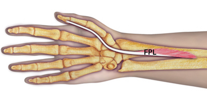
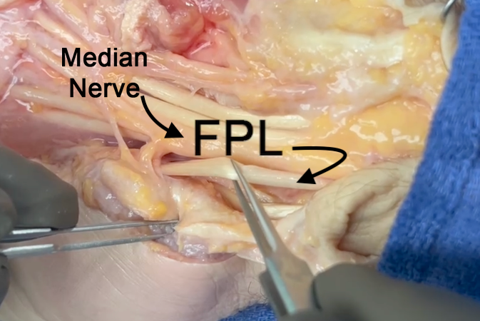
Flexor Pollicis Longus Anatomy
- Origin: Radius (anterior surface of middle 1/2) and adjacent interosseous membrane
Ulna (coronoid process, lateral border [variable])
Humerus (medial epicondyle [variable]) - Insertion: Thumb (base of distal phalanx, palmar surface)
- Innervation: Cervical root(s): C7 and C8
Nerve: median nerve (anterior interosseous branch) - The flexor tendon sheath of the FPL is an extension of the radial bursa.
- At the level of the thumb MP joint this synovial lined sheath becomes a fibro-osseous tunnel composed of the A1, A2, and oblique pulleys.
- In the thenar eminence the FBL is located between the superficial and deep heads of the flexor pollicis brevis.
- The long and short vinculum provide the blood supply to the FPL tendon.
- The oblique pulley which is an extension of the insertion of the adductor pollicis which is located superficial to the diaphysis of the proximal phalanx.
Diagrams & Photos
Key Points
- The princeps policies artery branches into the ulnar and radial digital arteries of the thumb. The thumb radial digital artery passes dorsal to the flexor pollicis longus (FPL) to reach its mid-lateral position on the radial side of the thumb.
- The radial digital nerve passes superficial to the FPL before reaching its mid-lateral position.
- During ORIF of distal radius fractures, the origin of the FPL muscle can be release to allow the FPL muscle and tendon to be moved ulnarly with the pronator quadratus to expose the volar distal radius.

