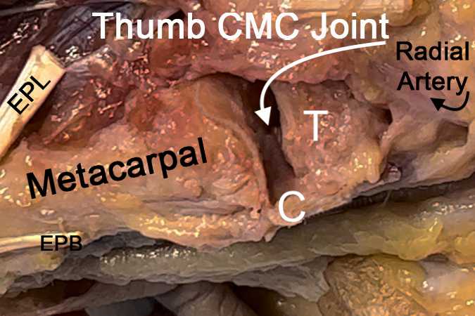
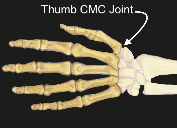
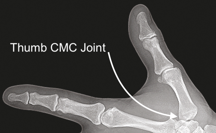
Thumb CMC (Basilar) Joint Anatomy
The Thumb CMC Joint provides an articulation between:
- First Metacarpal: The primary bone of the thumb connecting to the carpals.
- Trapezium: One of the carpal bones.
Ligaments:
- Anterior Oblique Ligament: Primarily responsible for stabilizing the CMC joint.
Tendons Crossing the Thumb CMC Joint:
- Abductor Pollicis Longus (APL) and Extensor Pollicis Brevis (EPB): Tendons that cross and influence movement at the CMC joint.
Joint Type:
- The thumb carpometacarpal (CMC) joint is a saddle joint with articular surfaces that are convex in one plane and concave in another. This saddle-like joint design allows motion in several planes, including the flexion/extension, abduction/abduction, and pronation/supination (rotational) planes.
- Synovial Joint
- Synovial joints are specialized structures that allow movement at bony articulations.
- Composed of a joint cavity lined by synovium containing bones lined with articular cartilage
- Structural components contain:
- Articular cartilage - enables low friction movement
- Ligament
- Joint capsule - fibrous tissue surrounding joint cavity
- Synovium - tissue lining non-cartilaginous portions of joint cavity and is composed of two layers, the intimal lining and the connective tissue sublining
- Synovial fluid - produced and regulated by the synovium
Diagrams & Photos
Key Points
- The CMC joint is integral for thumb mobility and can be affected by arthritis, especially osteoarthritis commonly termed as "basal joint arthritis."