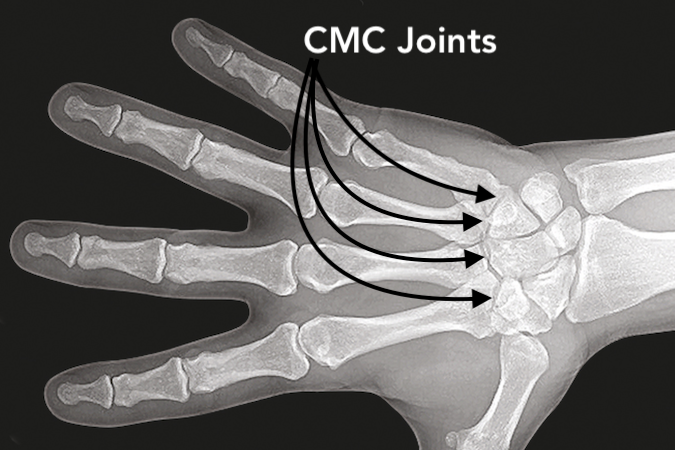
Carpometacarpal (CMC) Joints Exam
- The exam of the 2nd through 5th CMC joints begins by assessing the joints for deformity, bony tenderness, joint tenderness, and /or swelling.
- Next the 2nd through 5th CMC joints' passive and active range of motion should be noted.
- X-rays including AP, lateral and oblique views should be obtained to look for signs of fracture, joint subluxation, and/or joint dislocation.
- A CT scan maybe needed to evaluate the details of any fracture patterns.
Diagrams & Photos
Key Points
- The base of the 5th metacarpal has a convex facet articulates with a concave hamate facet to produce a saddle joint.
- The complexities of the 5th CMC joint make the evaluation of the fracture pattern and displacement of the baby Bennett fracture dislocation difficult.
- Dissections of the 2nd through 5th CMC joints have shown minimal degenerative changes in these joints. The OA findings range from 6% to 9%.
- Imaging of this joint frequently needs to be augmented with a 60 degree oblique X-ray view and/or CT scanning.
