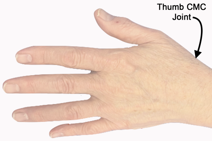
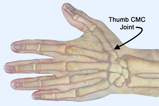
Thumb CMC (Basilar) Joint Exam
When examining an acute laceration of the thumb for signs of CMC joint injury, the goal is to determine if the laceration entered the joint, if the laceration damaged collateral ligaments and/or volar plate, and finally if the laceration damaged the joint cartilage.
Observation of the joint alone may be the only part of the exam that can be undertaken in the acute situation. Palpation and/or stressing the joint may cause severe pain. Therefore, the complete examination of the exposed joint surfaces and the ligamentous structures may require sterile technique and an anesthetized patient.
When examining a laceration associated with a joint, the first step is inspection of the laceration and observing its proximity to any nearby joint. Next, the examiner should assess the depth of the laceration, any visible joint deformity or abnormal angulation, and the laceration’s proximity to the joint cavity. With a severe laceration, such as a partial amputation, the inspection may immediately reveal that the joint is open and damaged. When the depth of the laceration is difficult to assess, the examiner may need to perform a surgical assessment under anesthesia to determine the open or closed status of the joint, the status of the joint’s ligaments, cartilage, and stability.
While the patient with a laceration may be able to demonstrate some active range of motion of the involved joints a complete assessment of active or passive range of motion may also require local, regional, or general anesthesia.
Additionally, while a simple laceration in the proximity of a joint may allow for gentle evaluation of the integrity of the collateral ligaments and volar plate by stress testing, a more significant laceration associated with the joint may be too painful to allow stress testing until anesthesia has been provided.
When the examiner inspects the joint, the uninjured or uninvolved opposite joint should always be viewed and used as a “normal” baseline.
Positive Exam
A joint exam associated with a laceration will be considered positive when there is clear evidence that the joint is open secondary to the laceration, if there is evidence of ligamentous injury, cartilage damage, or gross joint instability.
Negative Exam
A joint exam associated with a laceration will be considered negative when the joint is closed despite the nearby laceration and the joint is moving normally with no signs of instability or cartilage injury.
- If the patient’s laceration is unilateral, the clinician should always assess the asymptomatic “normal” side and use these findings as a baseline for determining the status of the injured joint.
- The injured “normal” side should be examined for signs of presence of hyperligamentous laxity. Congenital hyperligamentous laxity may explain joint instability that is not related to joint injury.
- The laceration may require examination with sterile technique under anesthesia before the examiner can make an absolutely accurate assessment of the joint’s involvement with the laceration and the overall integrity of the joint.
- The thumb CMC joint may be subluxed dorsally due to ligamentous laxity, injury, or arthritis.
- The CMC grind (compression) test can be used to demonstrate CMC crepitus and/or tenderness associated with thumb CMC joint arthritis.

