Distal Phalanx Anatomy
- The distal phalanges are very small terminal bones of the fingers and plays a crucial role in fine motor skills.
- The base of the distal phalanx, which is part of the DIP joint is covered with articular cartilage.
- The middle phalanx has three basic parts: The distal tuft the shaft or diaphysis, and the base.
- The concave proximal ends of the distal phalanx articulate with the middle phalanx.
- The proximal end of the 2nd, 3rd, 4th, and 5th distal phalanges articulate with the head of the 2nd, 3rd, 4th, and 5th middle phalanges.
- Dorsally the terminal extensor tendon inserts into a dorsal tubercle at the base of the distal phalanx.
- Palmarly the FDP tendon inserts into the palmar aspect of the base of the distal phalanx.
- The FDP inserts slightly volar and distal to the attachment of the volar plate to the base of the distal phalanx.
Diagrams & Photos
-
 Distal phalanx 16, 17, 18 & 19
 -
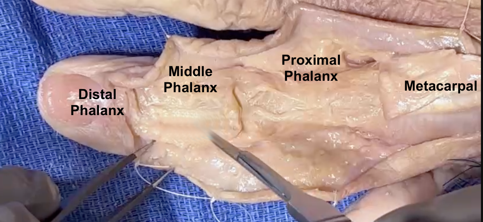
 Normal X-ray Image of distal phalanx, middle phalanx, proximal phalanx, and metacarpals.
 -
Key Points
- Numerous fascial bands attached in the skin of the pulp to the distal phalanx.
- An abscess (a felon) can develop in the fascial compartments of the pulp.
- The fingernail is firmly attached to the dorsum of the distal phalanx.
- The Landsmeer ligaments, specifically the Oblique Retinacular Ligament (ORL) attach to the dorsal proximal distal phalanx. This ligament helps synchronize PIP and DIP joint motion.
- In the growing child there is an epiphyseal plate in the base of the middle phalanx.
- The distal phalanges are fractured frequently.
- Distal phalanx fractures are often open fractures.