Index Extensor Tendon (Combined EDC & EIP)Anatomy
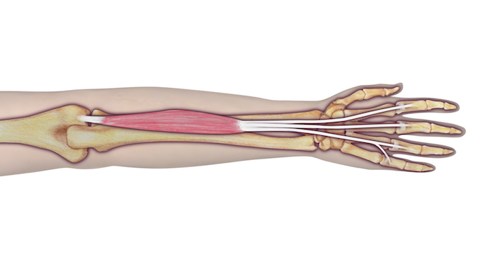
EDC:
- Origin:
- Humerus (lateral epicondyle via common extensor tendon)
- Intermuscular septum
- Antebrachial fascia
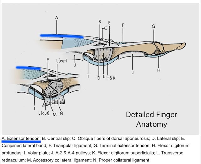
- Insertion: Four tendons to digits 2-5 (via the extensor expansion, to dorsum of middle and distal phalanges; one tendon to each finger)
- Innervation: Cervical root(s): C7 and C8Nerve: radial nerve (posterior interosseous branch)
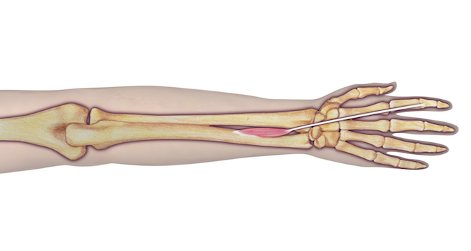
EIP:
- Origin: Ulna (posterior surface of shaft) Interosseous membrane
- Insertion: 2nd digit (via tendon of extensor digitorum into extensor hood)
- Innervation: Cervical root(s): C7 and C8 Nerve: radial nerve (posterior interosseous branch)
Diagrams & Photos
Key Points
- EDC extends the index, long, ring, and little fingers simultaneously.
- The EIP is ulnar and deep to the EDC in section 9 area.
- The Extensor Indicis Proprius (EIP) as a true proprius tendon provides independent extension of the the index MP joint.
- The EIP is a secondary abductor of the index MP joint.


