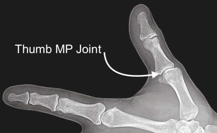
Thumb MP Joint Anatomy
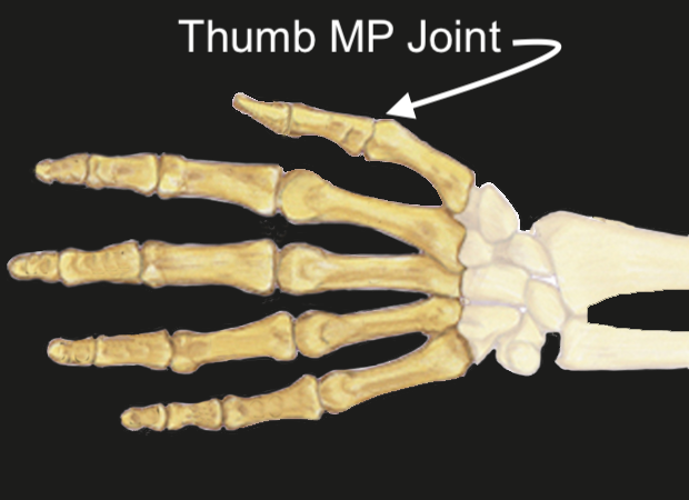
The Thumb MP Joint provides an articulation between:
- First Metacarpal: The primary bone connecting the thumb to the wrist.
- Proximal Phalanx: The bone of the thumb located between the metacarpophalangeal (MCP) joint and the IP joint.
Ligaments:
Ligaments:
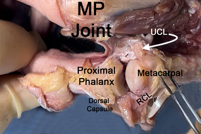
- Collateral Ligaments: These stabilize the joint from side-to-side movements.
- Volar Plate: Enhances stability on the palmar side.
Tendons crossing the Thumb MP Joint:
- Flexor Pollicis Brevis (FPB): Flexes the MP joint.
- Extensor Pollicis Brevis (EPB): Extends the MP joint.
Joint Type:
- The metacarpophalangeal (MP) joint of the thumb is a multiaxial condyloid joint. This ellipsoid-shaped joint allow arcs of motion in the flexion/extension and abduction/adduction planes, and to a lesser degree, in the pronation/supination (rotational) plane. The shape of the finger metacarpal head accounts for the “cam effect,” which allows abduction and adduction when the joint is in extension and little motion in the abduction/abduction arc when the MP joint is flexed. This explains the need to test MP joint collateral ligament stability in flexion.
- Synovial joint
- Synovial joints are specialized structures that allow movement at bony articulations.
- Composed of a joint cavity lined by synovium containing bones lined with articular cartilage
- Structural components contain:
- Articular cartilage - enables low friction movement
- Ligament
- Joint capsule - Fibrous tissue surrounding joint cavity
- Synovium - Tissue lining non-cartilaginous portions of joint cavity and is composed of two layers, the intimal lining and the connective tissue sublining
- Synovial fluid - Produced and regulated by the synovium
Diagrams & Photos
Key Points
- The MP joint is essential for thumb opposition and can be affected by conditions like arthritis or trauma.