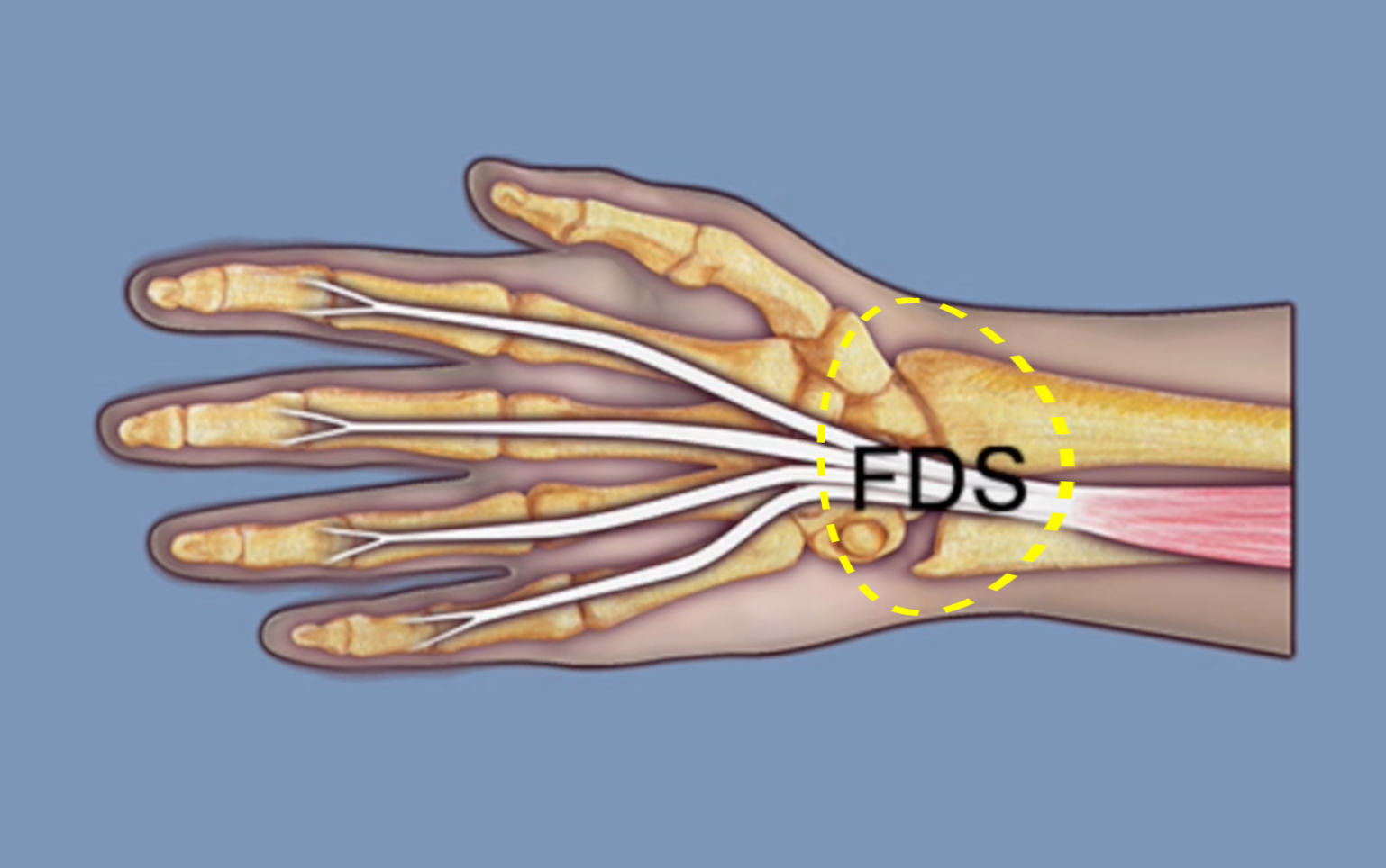
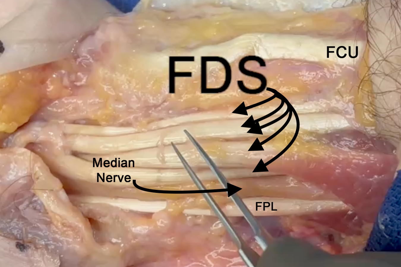
Flexor Digitorum Superficialis Anatomy
Flexor Digitorum Sublimis (FDS)
- Origin: Humeral-ulnar head: Humerus (medial epicondyle via the common flexor tendon), ulnar collateral ligament (UCL) of the elbow joint, ulna (coronoid process, medial side), and intermuscular septa. Radial Head: Radius (oblique line on anterior surface).
- Insertion: Four tendons arranged in two pairs: Superficial pair: Long and ring fingers, and deep Pair: Index and little fingers. All FDS tendons insert into the proximal middle phalanx.
- Innervation: Cervical root(s): C8–T1; Nerve: median nerve.
- In the proximal forearm the median nerve is on the dorsal surface of the FDS.
- The proximal flexor digitorum superficialis (FDS) has ulnar and radial heads.
- Proximally FDS muscle gets blood supply from the radial and ulnar arteries.
Diagrams & Photos
Key Points
- The flexor digitorum superficialis (FDS) is an extrinsic hand muscle that originates in the volar forearm.
- FDS II, III, IV, & V are median nerve innervated.
- The flexors also get nutrients from synovial fluid, local vessels, and in the finger from the vincula blood supply.
- There are two vinculum longus and two vinculum brevis with one longus and one brevis going to the FDS.
- At the Chiasma of Camper over the middle of the proximal phalanx the FDS splits into two slips which twist dorsally to reach their insertion sidtes at the base of the middle phalanx.

