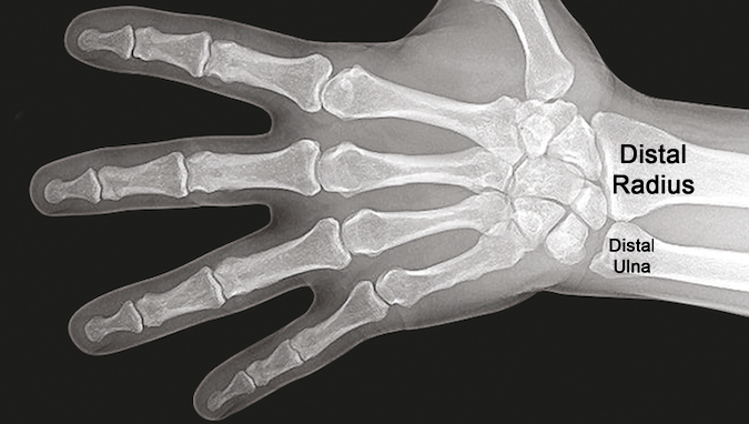
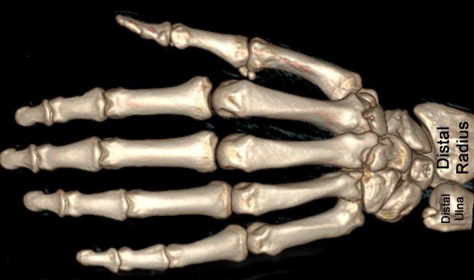
Distal Radius Exam
- When examining the distal radius look for deformity, swelling, loss of radiocarpal and radioulnar joint motion.
- Palpate for tenderness and crepitus.
- X-ray evaluation is typically needed.
- Evaluate the anatomy and bony alignment by measuring radial inclination, radial tilt, and radial length.
- The measurements done with an AP view and a facet lateral view which is taken with an X-ray beam inclined 10-15°.
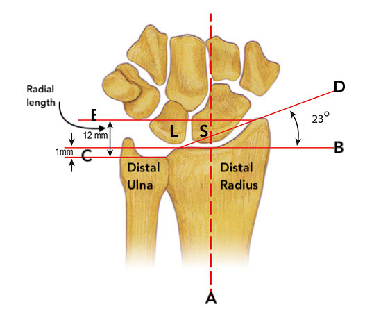
- Distal radius has a radial inclination or slope averaging 23°.
- The inclination drawing of the distal radius shows the lines used to calculate radial inclination. Line A is the longitudinal access of the radius. Line B is a line through the subchondral bone of the distal radius. This line is drawn perpendicular to line A. Line C goes through the subchondral bone of the distal ulna and is also perpendicular to line A. Line D is a line tangent to distal radius joint surface. The angle between line B and line D is the angle of inclination which averages 23 degrees. This drawing also shows a technique for measuring radial length using lines E and C.
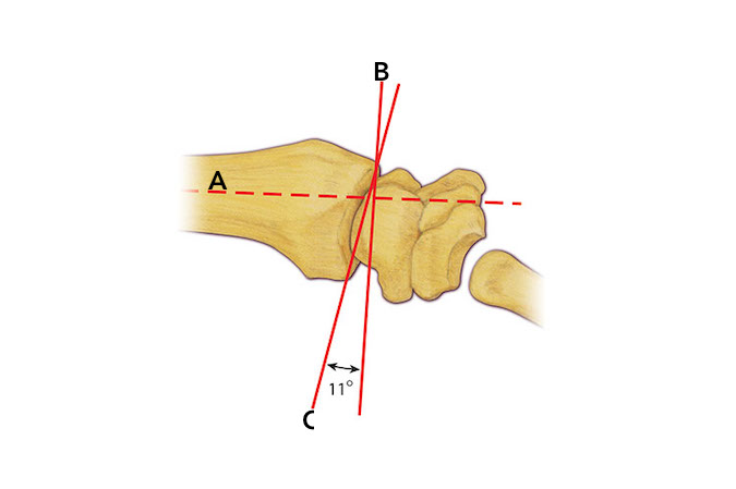
- Distal radius tilts palmarly (volarly) on average 11°
- The distal radius tilt is measured on a lateral X-ray. Line A is the longitudinal access of the radius. Line B is a line through the dorsal lip of the distal radius. This line is also perpendicular to line A. Line C is a line tangent to distal radius joint surface. The angle between line B and line C is the tilt of the distal radius which averages 11 degrees.
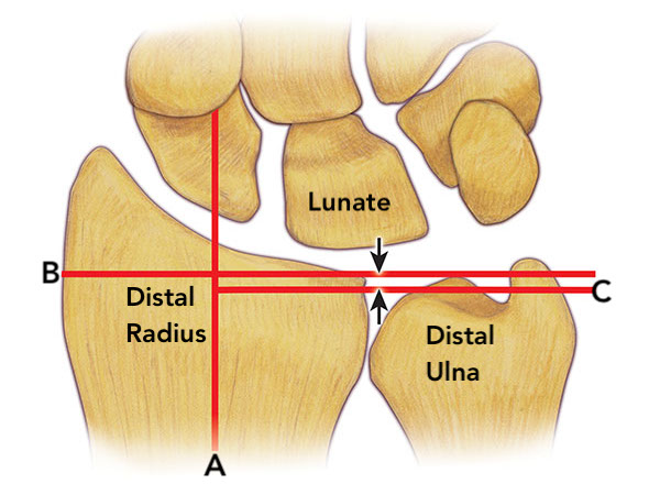
- Ulnar variance is used to measure relative lengths of radius and ulna on AP view. Ulnar variance is variable but averages 1 mm negative. This means the distal ulnar head is 1 mm shorter than the subchondral bone of lunate facet of the radius.
- Variance drawing of the distal radius, distal ulna, and carpus shows the lines used to calculate ulnar variance. Line A is the longitudinal access of the radius. Line B is a line through the subchondral bone of the distal radius. This line is drawn perpendicular to line A. Line C goes through the subchondral bone of the distal ulna and is also perpendicular to line A. The distance between the two arrowheads measured in millimeters is called the ulnar variance. When the ulna is shorter than the radius the variance is negative. The drawing depicts a negative variance of 1mm which is the average variance.
Diagrams & Photos
Key Points
- When evaluating fractures of the distal radius look for step-offs and/or gaps in the articular surface of the distal radius.
- Distal radius fracture evaluation may also require CT scans to properly visualize fragments, angulations, and degree of fragment displacement.
