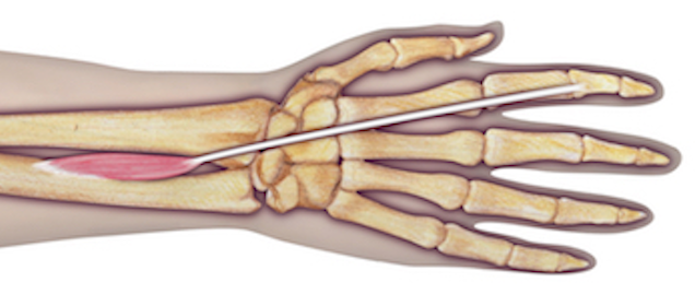
Extensor Indicis Proprius (EIP) Anatomy
Origin: Ulna (posterior surface of shaft) Interosseous membrane
Insertion: 2nd digit (via tendon of extensor digitorum into extensor hood)
Innervation: Cervical root(s): C7 and C8 Nerve: radial nerve (posterior interosseous branch)
Diagrams & Photos
Videos
EIP Anatomy Video
Key Points
- The EIP is ulnar and deep to the EDC in section 9 area.
- The EIP tendon is often transferred to the thumb to reconstruct the function of a chronic EPL tendon laceration.
